Severe Pumping Dysfunction

SEVERE HEART PUMPING DYSFUNCTION/ HEART FAILURE

Heart failure is a disorder in which the heart is unable to keep up the demands of the body, leading to reduced blood flow, backup of blood in the veins and lungs, or other changes that may further weaken or stiffen the heart.

Heart failure does not mean that the heart has stopped. It means the heart cannot keep up the work required to pump adequate blood to all parts of the body.

The function of the heart is to pump blood. The right side of the heart pumps blood from the veins into the lungs, while the left side pumps blood from the lungs through the arteries to the rest of the body.

Blood goes out of the heart when the heart muscle contracts (called systole) and comes into the heart when the heart muscle relaxes (called diastole).

Heart failure develops when the contracting or relaxing action of the heart is inadequate, typically because the heart muscle is weak, stiff, or both. Heart failure may be described as left-sided heart failure or right-sided heart failure.

Types of Heart Failure

  • Left-sided heart failure: Fluid may back up in the lungs, causing shortness of breath.
  • Right-sided heart failure: Fluid may back up into the abdomen, legs, and feet, causing swelling.
  • Systolic heart failure: The left ventricle cannot contract vigorously, indicating a pumping problem.
  • Heart failure with preserved ejection fraction: The left ventricle cannot relax or fill fully, indicating a filling problem.

Types of Heart Failure

Heart failure is classified by the ejection fraction (EF), which is the percentage of blood pumped out by the heart with each beat. A normal left ventricle ejects about 55 to 60% of blood.

The heart contracts less forcefully and pumps out a lower percentage of the blood that is returned to it. As a result, more blood remains in the heart. Blood then accumulates in the lungs, veins, or both.

The heart is stiff and does not relax normally after contracting, which impairs its ability to fill with blood. The stiff heart compensates for its poor filling by pumping out an even higher proportion of the blood than it normally does.

Eventually, as in systolic heart failure, the blood returning to the heart accumulates in the lungs or veins.

It is a newer concept that includes people whose ejection fraction is somewhere between preserved and reduced ejection fraction.

Types of Heart Failure

New York Heart Association Classification Scale on Heart Failure

Class I Heart Failure

There are no heart failure symptoms.

Class II Heart Failure

Everyday activities can be done without difficulty, but exertion causes shortness of breath or fatigue.

Class III Heart Failure

It is difficult to complete everyday activities.

Class IV Heart Failure

Shortness of breath occurs even at rest. This category includes the most severe heart failure.

severe-pumping-dysfunction
Heart Failure Causes Signs Diagnosis Prevention and Complications

  • High blood pressure
  • Coronary artery disease:The blood flowing to your heart is blocked or less than normal.
  • Cardiomyopathy:When the heart muscle is damaged, the heart cannot pump blood normally.
  • Heart valve problems:When heart valves do not open or close properly or become leaky, the heart has to pump harder to move blood through the body.
  • Congenital heart defect
  • Abnormal heart rhythms (arrhythmias)
  • Other diseases:Diabetes, HIV, overactive or underactive thyroid, or buildup of iron or protein.
  • Alcohol use
  • Sleep apnea
  • Smoking or using tobacco
  • Obesity
  • Viruses

  • Shortness of breath
  • Tiredness
  • Weakness
  • Swelling in feet, ankles, legs, or abdomen
  • Cough or wheezing
  • Dizziness
  • Confusion
  • Nausea
  • Lack of appetite

  • Blood tests
  • Electrocardiogram (EKG)
  • Chest X-ray
  • Echocardiogram (ECG)
  • Exercise test
  • Heart catheterization
  • CT scan
  • MRI
  • Coronary angiogram
  • Stress test

Preventing heart failure involves treating the disorders that cause heart failure:

  • High blood pressure
  • Obesity
  • Obstructive sleep apnea
  • Blockage of the coronary artery
  • Heart valve disorders
  • Abnormal heart rhythms
  • Alcoholism
  • Anemia
  • Thyroid disorders

  • Kidney damage or failure
  • Liver damage
Drugs for heart failure

Drugs for heart failure

Drugs to relive symptoms:

Diuretics, Nitrates, Digoxin

Drugs the helps to improve survival:

Angiotensin converting enzyme inhibitors, Beta blockers, Aldosteron antagonist, Angiotensin II receptor blockers, Angiotensin receptor inhibitors, Sodium glucose co-transporter 2 inhibitors, sinus node inhibitors.

The type of drug used depends on the type of heart failure. In systolic heart failure all the drug classes are helpful. In diastolic heart failure only ACE inhibitors, ARBs, Aldosterone antagonists and beta-blockers are typically used.

The following surgeries and devises may be required for some patients with heart failure

Heart bypass surgery

Heart bypass surgery creates a new route, called a bypass, for blood and oxygen to go around a blockage to reach your heart. The patient will be given general anesthesia. Once the patient is unconscious the heart surgeon will make an 8 to 10-inch (20.5 to 25.5 cm) surgical cut in the middle of your chest. Your breastbone will be separated to create an opening. This allows your surgeon to see your heart and aorta, the main blood vessel leading from the heart to the rest of your body.

Most people who have coronary bypass surgery are connected to a heart-lung bypass machine, or bypass pump. Your heart is stopped while you are connected to this machine. This machine does the work of your heart and lungs while your heart is stopped for the surgery. The machine adds oxygen to your blood, moves blood through your body, and removes carbon dioxide.

  • Infection
  • Heart attack
  • Stroke
  • Kidney failure
  • Lung failure
  • Bleeding

  • Eating a heart-healthy diet
  • Getting regular exercise
  • Treating high blood pressure and diabetes
Pre Procedural Imaging
Heart Valve Surgery

Heart Valve Surgery

Heart valve surgery is used to repair or replace diseased heart valves. Blood that flows between different chambers of your heart must flow through a heart valve. Blood that flows out of your heart into large arteries must also flow through a heart valve.

There are 4 valves in your heart:

  • Aortic valve
  • Mitral valve
  • Tricuspid valve
  • Pulmonic valve

Aortic valve and mitral valve are the most common valves to be repaired. Rarely, tricuspid and pulmonic valves are replaced or repaired.

Before surgery, you will receive general anesthesia.

In open-heart surgery, the surgeon makes a large surgical cut in your breastbone to reach the heart and aorta. You are connected to a heart-lung bypass machine if your heart is stopped while connected to this machine. This machine does the work of your heart, providing oxygen and removing carbon dioxide.

Minimally invasive valve surgery is done through much smaller cuts than open surgery, or through a catheter inserted through the skin.

Several Different Techniques Are Used

  • Percutaneous surgery
  • Robot surgery

Ring annuloplasty:Repairs the ring-like part around the valve by sewing a ring of plastic, cloth, or tissue around the valve.

Valve repair: The surgeon trims, shapes, or rebuilds one or more of the leaflets of the valve. The leaflets are flaps that open and close the valve. Valve repair is best for the mitral and tricuspid valves. The aortic valve is usually not repaired.

Types of Valves

Mechanical:Manmade materials such as metals or ceramics.

Biological:Made of human or animal tissues. These usually last 10 to 15 years.

Heart Pacemaker

A pacemaker is a small, battery-operated device. This device senses when your heart is beating irregularly or too slowly.

Pacemakers weigh as little as 1 ounce (28 grams). Most pacemakers have 2 parts:

  • The generator:Contains the battery and the information to control the heartbeat.
  • The leads:Wires that connect the heart to the generator and carry the electrical messages to the heart.

A pacemaker is implanted under the skin. This procedure takes about 1 hour in most cases.

You will be awake during the procedure.

A small incision is made on the left side of the chest below your collarbone. A pacemaker generator is then placed under the skin at this location.

Using live X-ray guidance, the doctor places the leads through the cuts into the veins and then into the heart. These leads are connected to the generator.

Types of Pacemakers

  • Transcutaneous pacemaker
  • Transvenous pacemaker

When your heart beats too slowly, your body and brain may not get enough oxygen.

Symptoms May Include

  • Light headedness
  • Tiredness
  • Fainting spells
  • Shortness of breath

  • Abnormal heart rhythms
  • Bleeding
  • Punctured lung
  • Infection
  • Puncture of the heart, which can lead to bleeding around the heart

Pacemakers can help keep your heart rhythm and heart rate at a safe level. The pacemaker battery usually lasts about 6 to 15 years. Your provider will check the battery regularly and replace it when necessary.

Heart Pacemaker
Implantable Cardioverter Defibrillator

Implantable Cardioverter-Defibrillator

An ICD is a device that detects any life-threatening rapid heartbeat. If an arrhythmia occurs, the ICD quickly sends an electrical shock to the heart. The shock changes the rhythm back to normal. This is called defibrillation.

The ICD is made of 3 parts:

  • The pulse generator contains the battery and electrical circuit that reads the electrical activity of your heart.
  • Your ICD has 1, 2, or 3 electrodes. Most ICDs have a built-in pacemaker.
  • There is a special kind of ICD called a subcutaneous ICD. This device has a lead that is placed in the tissue to the left of the breastbone rather than in the heart. This type of ICD cannot also be a pacemaker.

The surgeon places the electrode into a vein and then into your heart. This is done using a special X-ray to see inside your chest.

The surgeon then connects the electrodes to the pulse generator and pacemaker. This procedure usually takes 2 to 3 hours.

Why the Procedure Is Performed

  • Episodes of abnormal heart rhythms
  • Congenital heart problems or genetic health conditions

  • Blood clots that may travel to the lungs
  • Breathing problems
  • Heart attack or stroke
  • Allergic reaction
  • Infection

Your ICD will constantly monitor your heartbeat to make sure it remains steady. It will deliver a shock to the heart when it senses a life-threatening rhythm. Most of these devices can also work as a pacemaker.

Severe heart failure occurs when treatments no longer work. Certain treatments may be used when a person is waiting for, or instead of, a heart transplant.

  • Intra-aortic balloon pump
  • Ventricular assist device
  • Total artificial heart

Ventricular Assist Device

Ventricular assist devices (VADs) help your heart pump blood from one of the main pumping chambers to the rest of your body or to the other side of the heart. These pumps are implanted in your body. In most cases, they are connected to machinery outside your body.

A ventricular assist device has 3 parts:

  • Pump:The pump weighs 1 to 2 pounds (0.5 to 1 kilogram). It is placed inside or outside of your belly.
  • An electronic controller:The controller is like a small computer that controls how the pump works.
  • Batteries or another power source:The batteries are carried outside your body. They are connected to the pump with a cable that goes into your belly.
Ventricular Assist Device
Ventricular Assist Device Surgery

During Surgery

The heart surgeon opens the middle of your chest with a surgical cut and then separates your breastbone. This allows access to your heart.

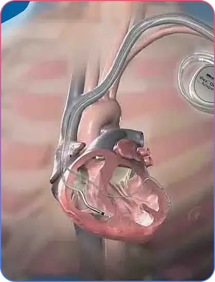
Depending on the pump used, the surgeon will make space for the pump under your skin and tissue in the upper part of your belly wall.

The surgeon will then place the pump in this space.

A tube will connect the pump to your heart. Another tube will connect the pump to your aorta or one of your other major arteries. Another tube will be passed through your skin to connect the pump to the controller and batteries.

Surgery most often lasts 4 to 6 hours.

There are other types of VADs (called percutaneous ventricular assist devices) which can be placed with less invasive techniques to help the left or right ventricle. However, these typically cannot provide as much flow (support) as the surgically implanted ones.

Why the Procedure Is Performed

You may need a VAD if you have severe heart failure that cannot be controlled with medicine, pacing devices, or other treatments.

You may get this device while you are on a waiting list for a heart transplant. Some people who get a VAD are very ill and may already be on a heart-lung support machine.

  • Blood clots in the legs that may travel to the lungs
  • Blood clots that form in the device and can travel to other parts of the body
  • Breathing problems
  • Heart attack or stroke
  • Allergic reactions
  • Infections
  • Bleeding

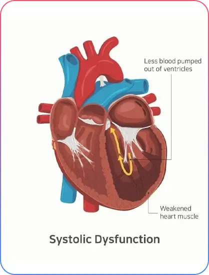
A VAD may help people who have heart failure live longer. It may also help improve patients’ quality of life.

Heart failure can suddenly worsen due to:

  • Ischemia (lack of blood flow to the heart muscle)
  • Eating high-salt foods
  • Heart attack
  • Infections or other illnesses
  • Not taking medicines correctly
  • Abnormal heart rhythms

  • Increased cough or phlegm
  • Sudden weight gain or swelling
  • Weakness

Go to the emergency room if you have symptoms like:

  • You faint
  • Fast and irregular heartbeat

CHF; congestive heart failure; left-sided heart failure; right-sided heart failure; cardiomyopathy; heart failure.