Chronic Total Occlusions

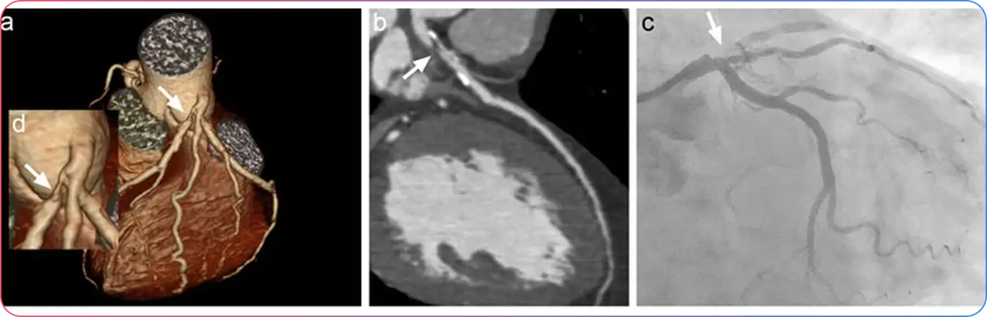
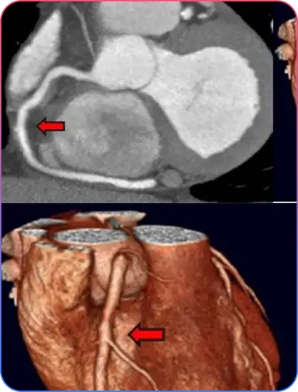
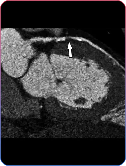
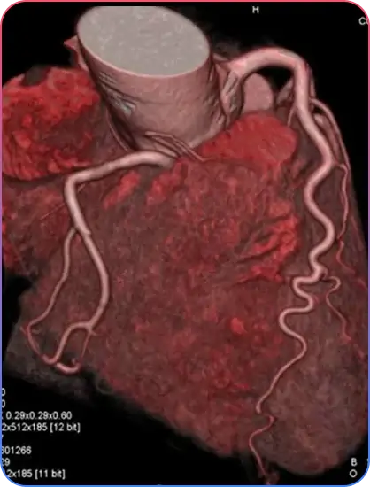
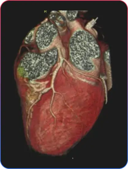
COMPUTED TOMOGRAPHY CORONARY ANGIOGRAM

Computed tomography (CT) coronary angiogram is an effective imaging test used to identify the plaque depositions in the arteries associated with the heart. This imaging test does not use any type of catheter insertion to the heart. The CT coronary angiogram uses the powerful X-ray equipment to produce pictures of the blood vessels and the heart. This technique is very safe and noninvasive.

CT coronary angiogram is a very effective way to identify various heart problems at an early stage. It is useful, especially, to diagnose atherosclerosis, even before any symptoms are observed. Plaque is made of various substances such as fat, cholesterol and calcium that deposit along the inner lining of the arteries that reduce or completely block blood flow.

Patients undergoing this scan receive an iodine-containing contrast material as an intravenous (IV) injection to ensure the best possible images of the heart blood vessels. The images generated during a CT scan can be reformatted to create three-dimensional (3D) images that may be viewed on a monitor, printed on film or by a 3D printer, or transferred to electronic media.

  • Blocked or narrowed arteries supplying blood to the heart
  • Chest pain (angina)
  • Irregular heart rate or arrhythmias
  • Inadequate supply of oxygen and blood to the heart
  • Difficulty in breathing
  • Dizziness
  • Fainting
  • Risk of heart attack

  • Abnormal anatomy of the coronary arteries
  • Low to intermediate risk atypical chest pain in the emergency department
  • Non-acute chest pain
  • New or worsening symptoms with a previous normal stress test result
  • Unclear or inconclusive stress test results
  • New onset heart failure with reduced heart function
  • Intermediate risk of coronary artery disease before cardiac surgery
  • Coronary artery bypass graft assessment
  • Assessment of calcium deposition in heart blood vessels and valves

  • Nausea
  • Vomiting
  • Diarrhea
  • Lightheadedness
  • Confusion
  • Constipation
  • Itching at the injection site
  • Allergic reactions to the contrast dye
  • Stroke
  • Arrhythmias or irregular heart rate
  • Kidney damage or kidney failure (rare)
  • Infections
  • Risk of blood clots
  • Hypotension
CT Angiogram Information
Aorto-Ostial Lesions

Before the procedure

  • Avoid eating at least 4 hours before the procedure.
  • Avoid consuming caffeine-induced beverages before the procedure.
  • Drinking water is allowed unless otherwise instructed by your doctor.
  • Remove all metal objects that may interfere with the procedure.
  • Inform the doctor about your past medical history, medications, and any allergies.
  • Inform your doctor about all current medications you are taking.
  • If you are diabetic, ask your doctor whether insulin should be taken before the procedure.
  • The patient's vital signs such as pulse rate, body temperature, blood pressure, oxygen levels, and breathing rate will be checked before the coronary angiogram.
  • Empty your urinary bladder before the procedure.

During the procedure

  • CT coronary angiogram is usually performed in the outpatient facility or the Radiology department.
  • The patient is asked to lie down on a padded table, which slides into a large hollow circular machine.
  • To examine the heart carefully, the doctor may give medications such as beta blockers to temporarily slow the heart rate.
  • The patient’s heart rate and rhythm are continuously monitored during the procedure using electrodes attached to the body.
  • The doctor cleans the area (arm or hand) where the contrast dye will be inserted.
  • The contrast dye is administered intravenously (IV) to highlight the heart arteries and help identify problem areas clearly.
  • Before inserting the contrast dye, local anesthesia is given to numb the area.
  • The procedure usually takes approximately one hour.
  • The X-ray machine captures images of the patient from different angles.
  • The patient is advised not to move and may be asked to hold their breath at times to obtain clear images.
Pre Procedural Imaging
Aorto-Ostial Lesions

After the procedure

  • Electrodes are removed from the patient’s body after the procedure.
  • Hospitalization is usually not required because the procedure is non-invasive.
  • The patient is generally able to drive back home without assistance.
  • The patient is advised to drink plenty of fluids to help flush the contrast dye completely out of the body.

Results

  • Findings if the report is abnormal:
    • Plaque formation or atherosclerosis
    • Reduction in blood flow through the arteries
    • Reduced blood supply to the heart and surrounding blood vessels
    • Abnormalities in the structure and condition of the heart, such as congenital heart diseases
  • If the report is normal:
    • No abnormalities are detected
    • The heart is healthy and functioning normally
    • No obstructions or blockages are observed
    • The structure of the heart appears normal
Pre Procedural Imaging
Aorto-Ostial Lesions

Lifestyle modifications

  • Avoid smoking and alcohol consumption.
  • Eat healthy and nutritious food.
  • Maintain a healthy body weight.
  • Avoid foods high in saturated fats.
  • Manage stress effectively through relaxation and healthy lifestyle habits.
  • Engage in regular physical exercise as advised by your doctor.
  • Interventional or surgical heart procedures may be advised if required.