Intravascular
Intravascular Ultrasound (IVUS) is a medical imaging methodology, which uses an ultrasound probe that is inserted to the artery like a regular angioplasty catheter to provide an image from within the artery.
Used to see the inside blood vessels using soundwaves. A computer produces pictures of soft tissues. This technique allows physicians to see areas that they can’t see with X-rays
Cardiologists use IVUS to visualize exactly location of plaque, which helps determining whether stenting is possible, or whether a patient might require surgery.
IVUS are also used to assess stents that have already been inserted & whether they have expanded properly.


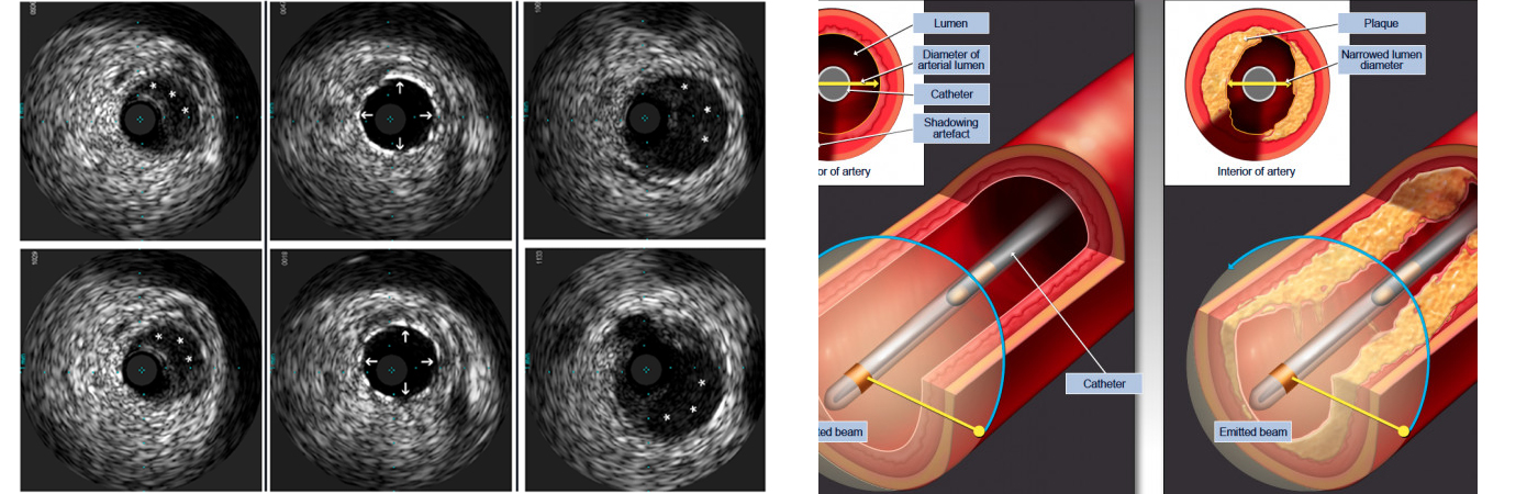
How does
IVUS work?
Transducer/ Probe emits the soundwaves
Why use
IVUS?
To view the artery from inside out
Preparing for procedure
Follow specific instructions given b your doctor or nurse about what you can and cannot eat or drink before the procedure.
Avoid medications such as Warfarin (a blood thinner) or Aspirin.
Inform if you have any allergies, especially x-ray dye, latex or rubber products or penicillin-type medications allergy.
Preparing for procedure
Please tell the doctor or nurses if you feel chest discomfort or any other symptoms during the procedure.


Post procedure Care
Rest as per your doctor’s suggestion
Inform immediately if any of the following symptoms are seen near incision site- pain, warmth bleeding, swelling, or a change in colour.
Contraindications
Bleeding disorder
Stroke
Arrythmias
Pregnancy
Inability of patient cooperation.

"Dr. Sharath Reddy is the best cardiologist in Hyderabad. We underwent the heart treatment under him. We are very happy with the treatment."
I would like to thank Dr Sharath Reddy and his team for the excellent cardiac care and treatment provided to me. I have been given all the information and advice to lead my life in a comfortable way after the procedure. I can’t thank you enough for all your time, hard work, and patience. I truly appreciate your caring dedication.
My brother had severe dyspnoea (shortness of breath) and it was difficult for him to walk for even very small distances. He had severe pedal oedema (feet swelling) and was not able to do routine activities on his own. But after TAVI procedure he is able to breathe well and walk comfortably for small distances without any difficulty. Within a short time after the TAVI procedure he is performing his routine own activities. We are grateful to the cardiac team at Medicover Hospitals.