Advanced Imaging
Cardiac imaging is a subspecialty of diagnostic radiology. A cardiac radiologist supervises or performs and then interprets medical images to diagnose diseases of the heart such as heart disease, leaky heart valves and defects in the size and shape of the heart.
A physician may recommend cardiac imaging to support a diagnosis of a heart condition.
Options
Echocardiography
An echocardiogram (echo) is a graphic outline of the heart's movement.


Cardiac Catherization/Angiogram
It is an invasive procedure, where a thin, long, flexible tube is inserted, via the arm or groin, and is guided to the blood vessels of your heart to evaluate and treat heart conditions.
CT Angiogram
It combines a CT scan with an injection of a special dye to produce pictures of blood vessels and tissues in a part of your body.


CAC Scanning
Measures the amount of calcium in the walls of the heart's arteries.
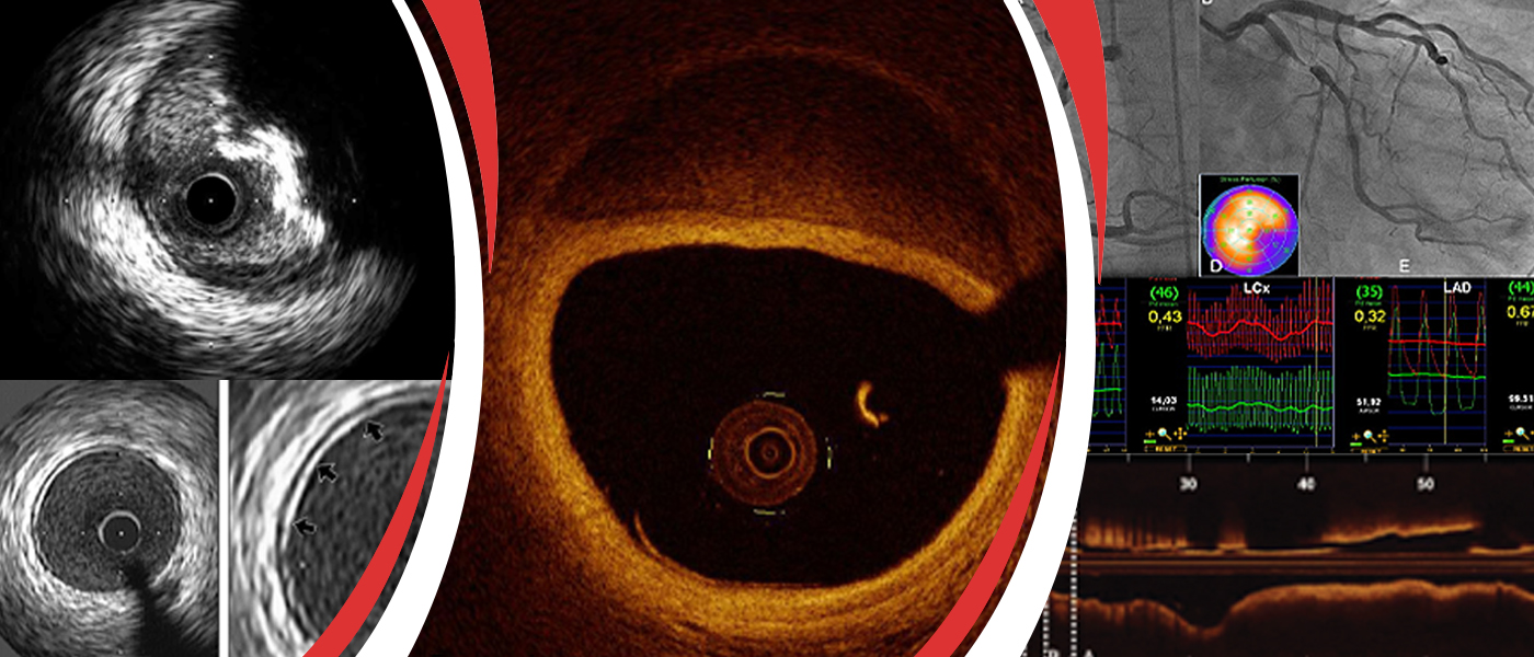
INTRAVASCULAR ULTRASOUND (IVUS)
Intravascular Ultrasound (IVUS) is a medical imaging methodology, which uses an ultrasound probe that is inserted to the artery like a regular angioplasty catheter to provide an image from within the artery. Used to see the inside blood vessels using soundwaves. A computer produces pictures of soft tissues.


Fractional Flow Reserve (FFR)
Fractional flow reserve (FFR) is a technique used in coronary catheterization to measure pressure differences across a coronary artery stenosis to determine if stenosis impedes oxygen delivery to the heart muscle.
Optical Coherence Tomography (OCT)
Optical Coherence Tomography (OCT) is a diagnostic procedure that uses near-infrared light to create images of the inside of the coronary arteries. The technique delivers very high-resolution images.

"Dr. Sharath Reddy is the best cardiologist in Hyderabad. We underwent the heart treatment under him. We are very happy with the treatment."
I would like to thank Dr Sharath Reddy and his team for the excellent cardiac care and treatment provided to me. I have been given all the information and advice to lead my life in a comfortable way after the procedure. I can’t thank you enough for all your time, hard work, and patience. I truly appreciate your caring dedication.
My brother had severe dyspnoea (shortness of breath) and it was difficult for him to walk for even very small distances. He had severe pedal oedema (feet swelling) and was not able to do routine activities on his own. But after TAVI procedure he is able to breathe well and walk comfortably for small distances without any difficulty. Within a short time after the TAVI procedure he is performing his routine own activities. We are grateful to the cardiac team at Medicover Hospitals.