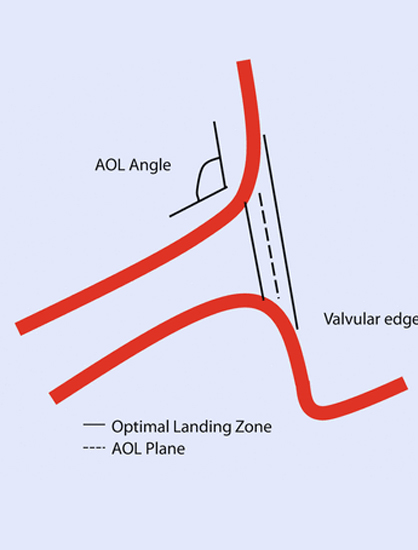
Ostial Lesions are described as coronary ostial stenosis, is the occlusion of coronary ostium located within the first 3 mm from the beginning of the coronary artery.


Causes
Atherosclerosis

Diagnosis
- Angiography

Types
Aorto-ostial lesions
Aorto-ostial leisions
- Aorto-ostial coronary lesions (AOL) are defined as a stenosis within 3 mm of the right coronary artery (RCA) or left main coronary artery (LMCA)
- Aorto-ostial lesions commonly have a unique three-dimensional funnel-shaped morphology with a variable angle of takeoff of the coronary artery from the aorta
- The majority of the AOL are accompanied by diffuse coronary artery disease and cases of isolated AOL occur predominantly in females
- Aorto-ostial lesions were examined postmortem in victims of acute myocardial infarction (AMI), sudden death without pathological evidence of AMI and violent death.


Pre-procedural imaging
- CCTA
- IVUS Intra-procedural imaging:
- Integrated IVUS on stent delivery system
- Fusion of IVUS and CCTA with angiographic imaging
- Devices for imaging the aorto-ostial plane
Diagnosis and treatment
- Diagnosis and treatment of these lesions is challenging and procedural success and clinical outcomes are inferior to non-ostial lesions.
- Diagnosis of AOL may be missed on coronary angiography when contrast media is injected distal to the lesion due to deep intubation of the catheter into the vessel.
- Intravascular ultrasound (IVUS) is a useful tool for detailed analysis of the AOL anatomy
- Various debulking strategies advocated for optimizing the outcome of angioplasty in rigid AOL included directional atherectomy, rotational atherectomy , excimer laser and cutting balloons.
- Introduction of bare metal stents reduced acute procedural complications and re-stenosis rate compared to balloon angioplasty
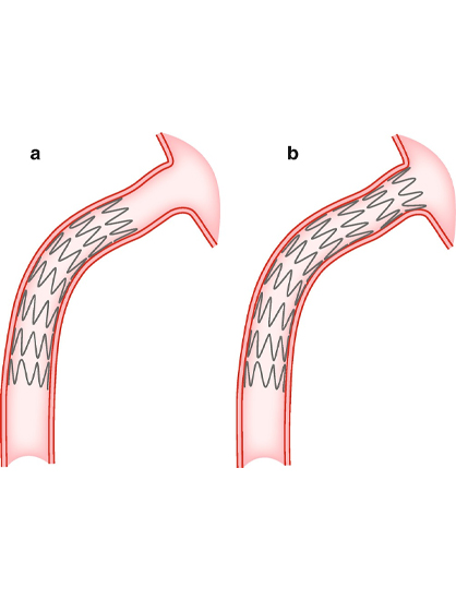
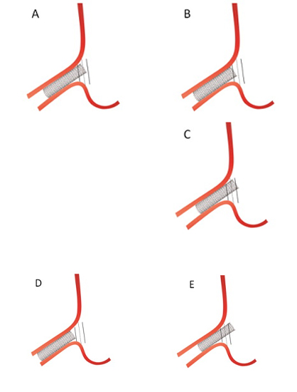
Implantation of short stents and excessive instrumentation following deployment may lead to stent dislodgement and embolization to the aorta - Direct contact between the guiding catheter and the proximal edge stent struts may lead to longitudinal stent deformation.


